The diagnosis of cavitation lesions is complicated by the fact that x-ray examination of the jawbones often appears normal . . . to the untrained eye. Considerable diagnostic experience is required to detect disorders that mimic cavitations, including variations of normal anatomy.
Why is this so? Osteonecrosis is a disease of the marrow spaces of bone and 40% to 50% of such bone must be destroyed before changes can be seen on x-rays. So, if your dentist or oral surgeon takes an x-ray and pronounces the film normal in spite of your symptoms, don’t necessarily believe it. X-rays may be interpreted as normal unless (1) there’s a significant amount of bony destruction or (2) the doctor is experienced in reading x-rays specifically for cavitations.
Although MRI (magnetic resonance imaging) is the imaging technique of choice for long bones, flat bones of the face are not imaged well with regular MRI scans. CT scans are also ineffective in locating most cavitations in the jawbones.
Figure 15: MRI STIR image. The cavitation is the larger white area on the right side of the picture.
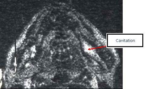
However, we’ve discovered that using the technique of MRI STIR imaging (Figure 15) is very effective and accurate in locating areas of bone marrow edema (swelling) and ischemia (areas of reduced oxygen). Both of these conditions can and do lead to the formation of cavitations.
Figure 16: Bone scan using tech 99 radioisotope. The cavitation is the darker area in the lower right front.
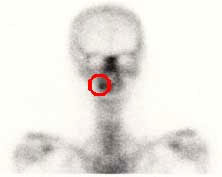
one scans using a radioactive isotope are somewhat helpful in locating cavitations but very difficult to interpret. Also, radiologists, not expecting these lesions in jawbones, often note the lesions in their radiology reports but interpret the results as normal.
The best, most effective method to locate cavitations is the Cavitat bone scanning device (Figure 17). This computer-based sonar imaging system was designed to aid the medical community with a detailed profile of the interior of bones. The Cavitat computer generates digitized two and three dimensional images of the interior of the jawbones from sound waves passed through the bones.
Figure 17: Cavitat scan.
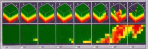
Because liquid is a near perfect conductor of sound waves, when these waves enter into voids or porosities in bone (areas that have compromised bone flow; i.e., cavitations), the sound waves slow down considerably, which produces images of the interior of the bony area being scanned. We’ve found the Cavitat results to be very accurate, especially when compared with patients’ panoramic x-rays. Our diagnostic results have improved dramatically. Most importantly, our surgical successes have soared since we began using this revolutionary device.
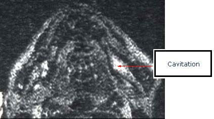
Therefore, since both MRI STIR imaging and ultrasound imaging (Cavitat) are so effective and accurate (Figure 18), since November of 2003 we’re been using both imaging techniques with most patients. Using both of these diagnostic tests have helped improve our diagnostic abilities and better yet, have improved our overall success rate in treating cavitations of the jaws.
Figure 18: MRI STIR image and Cavitat scan of the same area, both demonstrating a cavitation in the same area.
For patients experiencing pain, diagnosis is further improved through anesthetic confirmation or anesthetic blocking. By giving a local anesthetic injection (similar to having your dentist numb the jaw before he or she performs a dental procedure), pain in the jaws can be selectively turned-off, meaning the sense or feeling of pain can be chemically and temporarily eliminated. If the pain goes away after the injection, then we can be reasonably certain that there’s a problem in the anesthetized area, generating pain.
The only treatment available at this time to remove cavitational lesions is surgical removal. Some have attempted to inject homeopathic remedies or ozone into these areas of dead bone, but unfortunately, there’s no blood circulation within cavitational lesions, so any medications, drugs, or remedies can’t get into and permeate these lesions, let alone allow toxins and metabolic products to be removed. Homeopathic remedies certainly have their place in NICO treatment, especially in healing after surgical removal of the lesions themselves.
The surgery basically consists of making an incision, exposing the bony defects, and scraping them clean (termed debridement) to remove all unhealthy bone and other pathological problems like abscesses and cysts. It’s not sufficient to simply punch a hole in the bone and rinse the area out, like some doctors recommend. In fact, treating these expanding bony lesions in such a conservative fashion often makes the lesion and subsequent pain much worse.
After removing the dead bone and other pathological products, the goal in healing is bone regeneration. But first, if possible, we remove all predisposing and risk factors.
If you think you might have a NICO lesion, what can you do? First, find a doctor who understands this disease process; one who is trained in effectively diagnosing and treating these bony problems. Unfortunately, there are precious few such doctors in the world and very few in North America at this time.
If you're experiencing pain, don’t allow anyone to operate without first proving where your pain originates. This is done most effectively by closely evaluating x-rays and using diagnostic anesthetic injections to actually turn-off the suspected NICO areas to see if the pain is turned-off. There are characteristic referred pain patterns of NICO lesions and there are also characteristic responses to local anesthetic testing. Find a doctor who knows about these characteristic patterns and realize that most doctors who treat orofacial and TMJ pain know nothing about NICO lesions.
Be certain that the doctor obtains Cavitat scans, MRI STIR imaging, or both in the process of diagnosing your problem. Both of these imaging tests give us a view of the size and extent of cavitations and can also indicate if surgery is truly needed or not.
Keep watching this site as we have many new and exciting things soon to come out about NICO lesions. For more information about NICO lesions and other orofacial pain problems which are often misdiagnosed, see Dr. Shankland’s latest book, Face The Pain.
Dr. Shankland consults and treats NICO, orofacial, and TMJ patients. However, to be in compliance with and cooperate with the Ohio State Dental Board, potential patients must understand that currently, the diagnosis and treatment of osteocavitations is considered experimental and alternative. Further, if you would like to consult with Dr. Shankland concerning this disorder, you first must have a referral from a physician in order to be seen by Dr. Shankland. If you have any questions, please call Dr. Shankland’s office ( 614-794-0033 ) and ask for the NICO Information Packet to be sent to you free of charge. You can also consult with Dr. Shankland. If you’d like to know more about Dr. Shankland, click on Biography.